Showing posts with label Pictures. Show all posts

Thursday, February 21, 2013

Muscle and More

Though I have often come back to the topics of biology and Special Creation here, I don’t intend to have a teaching blog about cell biology or genetics. Websites dedicated to teaching various disciplines abound on the Internet, not to mention school courses that are available. I have wanted to be another voice speaking about the design of biology and how I believe God created us. But I would rather write generally about this topic than specifically for the most part, and also address other concerns.

The posts I have made about biology are mostly for adults, although I believe children could appreciate them. It is adults who determine what is taught to children, and I want parents to think about how biology and evolution are presented in the schools. But some of the other themes I have written about are disturbing. I guess we are used to encountering bad interspersed with the good in our lives, but my blog wouldn’t succeed that way as a consistent lesson in Creationism. So I want to give you some references for other websites which do teach it, so you can continue to learn specifically about this subject.

I just found a Catholic Creationist site for the Kolbe Center for the Study of Creation. It looks like there is plenty there to read, including Dr. Robert Bennett, "A summary of theistic evolution" .

Though Answers in Genesis is not Catholic based, it is also a Young Earth Creationist website. It gives insight as to how the Earth shows evidence of Divine Creation. It can be a reference point for many of your questions, since it is well organized and addresses geology and cosmology as well as biology. You may be surprised how much information is available that opposes the mainstream science “party line.”

Another website where you can appreciate biology is Creation Wiki. There are different categories in the main page that will lead to entry links. They have links you can follow to other subjects.  Creation Wiki doesn’t have as many entries as some other encyclopedias, so you may have to look further for more detail. But that should be true for any research you do. Different references can be complimentary and the more you search and read, the more you learn.

There is a website called creation.com which is an international ministry for the promotion of Creationism. They are involved in an interesting campaign called “Question Evolution.” I hope they will make inroads against the enforced teaching of totally materialistic, naturalistic evolution.

I also found a site with some interesting videos of biology, DNAtube . Unfortunately, as with many scientific biology sites, you have to step around evolutionist talk.

It’s kind of amazing how much biologists and other scientists protest Creationism. If Darwinian evolution is as much a fact as they say, it should be easily apparent. However, the opposite is more like it. Fabulous designs are everywhere, and it is an exercise in futility to try to explain cell biology as random globs of molecules.

Though others may want to suppress Creationists and have tried to do so in many ways, we still have the freedom to observe and describe biology as it really is. I wish you happy hunting and learning.

~~~~~~~~~~~~~~~~~~~~~~~~~~~~~~~~~~~

All the above said, I’d still like to show you another wonderful biological design. In a recent post I talked about the tiny bio-machines that produce energy for our body. The small molecule that carries this energy to other chemical reactions is called adenosine triphosphate (ah-DEN-oh-seen try-FAHS-fate). The commonly used nickname is ATP. The mechanism for muscle contraction is one of the many uses for ATP. Our muscles have muscle cells with muscle proteins inside. These have technical names, which you can learn if you are inclined. But you don’t have to know their labels to see the way they operate. An explanation of muscle is at the Wikipedia website HERE and the link to the picture is HERE.


Two types of proteins form strands that interact to pull and release so the muscle can move. One of these strands has a lever at the end which can flip when ATP combines with it. When the energy from ATP is released, the remaining part is called ADP (adenosine diphosphate). The “tri-” and “di-” before the phosphate indicate a section of the molecule was removed when energy was given off. ADP has to be eventually turned back into ATP by the mechanism given in the other post. A good image and description is by Jeff Sale (Source HERE© statement HERE).:


(Magnesium ion is gray against the red myosin head. ADP and phosphorus ion are dark at side of red myosin head. Actin filament goes across image at angle.)

It is a wonder, all that has to work in order for us to have life. I hope anyone with children will help them understand the wonders of biology.

Thursday, January 31, 2013

Beneath the Surface

I am adding another post to my blog about the subject of biology. This is not to confuse you but to hope you appreciate the vastly complicated interactions that must take place in order for life to occur and proceed. Evolutionists talk about fossils and bones, but beneath the surface of individual bacteria, plants and animals is a vast interacting world which must function in order to get life going and continuing.  I have a few pictures below to give you an idea of these activities.

Did the organized biochemistry of life happen by totally materialistic, naturalistic means through chance? On one hand, the current theory, called “neo-Darwinism” claims that natural selection, known as “survival of fittest,” is not by chance, but comes about by competition of those who reproduce. But on the other hand according to the same neo-Darwinism, any superiority of individuals depends on random mutation of their genes. They can't get away from the fact that materialistic evolution is ultimately based on chance.

Neo-Darwinism is not better and is actually less logical than the belief that God made species directly. I wonder sometimes what evolutionists are thinking. In fact, I’ve got to believe there are many scientists out there who are not saying much publicly but must seriously doubt whether totally materialistic, naturalistic evolution can be true.

That is why I am taking the time to show you some of the facts. Though I’m not an expert in cell biology, I have a background in biology (I graduated from two universities with a BS in animal science and a VMD in veterinary medicine). I have spent a lot of time reading scientific research on genetics, biochemistry and cell biology.

My last post was about the complexity of the cell’s energy-making system. But, it was actually only a part of that system. Another part is needed to process food we eat in order to get pieces of it to feed into this system. In humans, the process is known by a few names, one being the citrate cycle (CreationWiki description under cellular respiration HERE).

Chemical interactions between molecules are the underpinning of biological function. The molecules are made of atoms, with which most of us are familiar. The air we breathe is made of various atoms, including oxygen. These combinations of atoms are often described with terms we don’t recognize unless we study them, but we can still understand some concepts without knowing all the names of the molecules. There are more and more databases of information about biology and other disciplines available. Scientists add to this data from their discoveries and use it for further research. A lot of the information is accessible by Internet, which is a wonderful service to scientists and anyone else who wants to learn.

One of these databases is called KEGG, for Kyoto Encyclopedia of Genes and Genomes. I had a picture of part of the energy system from KEGG in my last post. Here I show an overview map (image 1 of this post, link HERE for source) of the biochemical activities of cells which KEGG has put in the database. (You can go to the link to see more and can also click on the picture here to get a larger view.) The paths contain biochemical reactions for various species.  For example, it describes how the body processes things like fats and proteins that we eat. These chemicals must have precise coordination of molecules for fitting together and producing the desired outcome in each species.

The map does not nearly cover all the biochemical reactions that exist. In fact, the current estimate for the number of species on Earth is over 11 million by Liz Osborn at Current Results website HERE. National Institutes of Health researchers did a study on the single-cell organisms of bacteria and what are called archaea (are-KEY-ah). The article was by Koonin and Wolf, "Genomics of bacteria and archaea," Nucleic Acids Research, 36, 21 (Dec. 1, 2008): 6688-6719. They found that “remarkable biochemical diversity is a hallmark of bacterial and archaeal biology.” So you can be sure there is much out there that is not yet on this map. These are the chemical data that the people who do the map know about and have inserted.

The citrate cycle is a small portion of biological metabolism. In the overall map, the cycle is named inside the small round blue line in about the middle near the bottom. If you go to the KEGG site and click on the “citrate cycle” name, it will bring you to a chart of the chemical reactions that happen in the cycle. I have a picture of the human citrate cycle that comes up, KEGG source linked HERE (image 2 of this post). The green boxes are links to further information about the human biochemical reactions. They show facts about the proteins called enzymes (EN-zimes) that work to change molecules within the cycle so they produce the necessary segments for energy production.

Don’t be concerned if these terms sound foreign to you. I’d just like you to get an idea of what goes on in our bodies. One of the green boxes shows the number for enzyme 4.2.1.3. This is a protein enzyme called aconitate hydratase (ah-CON-ih-tate HI-drah-tase). If you click the box at the KEGG website, you get information, with more links, about aconitate hydratase, entry EC:4.2.1.3.  The rectangular box here is what you would see (image 3).

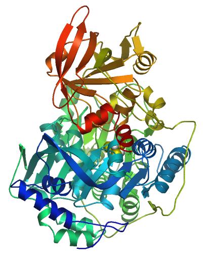
The box contains a picture of the protein. I want to show you a similar, larger picture from another database of the aconitate hydratase protein as they use it in models. The last picture here (image 4) is from Swiss Model Repository, entry Q99798. Swiss Model Repository is a database that shows the 3 dimensional models of proteins. The swirls and arrows show the types of folds that the amino acids make in order to form the protein so it does a specific job. This protein is made up of 780 amino acids which must be in a particular order (if you are interested in more details of the protein, you can get them at yet another database, Uniprot, entry Q99798). At the bottom of the third picture, there is a list of amino acids that make up the enzyme that we are talking about. (For information on amino acids, go to CreationWiki HERE.)  For purposes of saving space, I did not add the bottom section of the box, which lists the specific order of the DNA of the protein's gene.

If you have gotten this far, I appreciate that you have been interested enough to read about the complexity of cell biology. It is important because of what I’ve been talking about concerning the question of neo-Darwinian evolution and whether it actually happened. Though you may hear some scientists or their spokespersons say evolution is a fact, the possibility and reality of it is something you have to decide for yourself.

Thursday, January 24, 2013

Cell Biology

When you start learning biology, there are new terms. With computers, your job, and/or your school, you learn new things all the time, so you are already acquainted with the activity. So I hope you will be open to learning about cell biology. It is a fascinating subject, and well worth the time. Perhaps you already know biology, but I hope you will still look at what I have here.

There are an estimated 75 trillion cells in the human body (and trillions of atoms in each cell). From bacteria to humans the cell is the basic unit of biology, and its activities are called “metabolism.” The cell absorbs and stores food, then breaks it down and converts it into energy. It uses energy to reproduce DNA (genes) and assemble proteins among other things. The DNA is used as a code for the proteins  which in turn do the work of the cell and the body. For example, your muscles have muscle cells which contain muscle proteins that contract and relax. They are called by specific terms, but we don’t have to name everything to get the overall perspective.

One of the basic parts of the ongoing process of metabolism is where several groups of proteins are embedded in a membrane of of the cell. The membranes are in folds, and in humans the folds are parts of what are called mitochondria (mite-oh-CON-dree-ah). First I have a short video (less than 4 minutes) for you to see how one of the groups works to form the energy molecule called ATP. ATP has chemical qualities that give it the ability to use a chemical bond for activity needed in the cell as I described above. It changes to ADP when used up, then is re-cycled back to ATP by this complex. You may hear some terms you don’t understand, but just try to get the concept of this series of steps going on in most of your body’s cells.

The video is done by North Dakota State University which has done other animations you can see HERE if you want to learn more. As interesting as the above video is, the protein complexes are drawn rather simply. Below is a more detailed picture of a series of complexes needed to make the last one, ATP Synthase (SIN-thase), work. They are needed to produce the electrical gradient which is described in the video. The mitochondrial membrane is pictured between the complexes, with ATP Synthase at the far right.

The image (link HERE) comes from a database called Kyoto Encyclopedia of Genes and Genomes (KEGG). The boxes in the picture give information for various parts of the complex when you click on them at the KEGG website.

Now, please stick with me a little longer to get more of the perspective of how complicated this series is. In the picture, the last group of proteins on the right is ATP Synthase. On the bottom part, there are blue-colored sections. The darker blue have a β on them for beta subunit (beta is Greek for “B”). Proteins are made of yet smaller units called amino acids (ah-ME-no acids, Wikipedia entry HERE). These are repeating groups of atoms, including carbon, oxygen, nitrogen and hydrogen (C, O, N and H). Here is a picture of one of the 20 types of amino acids found in biology, called alanine (AL-ah-neen):

Carbon is also understood to be in the angles of the molecule image. The types of amino acids need to be in correct order, according to their size, electrical charge and other factors, so the proteins can fold into the shapes in which they function. It is similar to machine parts which need to have the right shapes to fit and move together. Now I have just one more picture. It is the list of amino acids in this one part of the whole complex, the beta (B) subunit of ATP Synthase as pictured above. This particular one (human) has 529 amino acids, as listed in another database called Uniprot, entry P06576. Each letter stands for an amino acid, such as A for alanine (letters can stand for different things depending on their context):


All the proteins parts of this entire complex are made of various arrangements of the amino acids. Many are at least 100 amino acids and some are many more, as you have seen. These need to be constructed and put together within most of the cells that we have. There are many such complexes within each cell.

Thank you for bearing with me to learn about this part of cell metabolism. Remember, this is only a very small fraction of the complexity in the cells. Even small organisms need energy systems such as these to put together their own genes and proteins. Please think about whether this could have come about by chance, no matter what the time frame. After all, a computer would not form on its own, no matter how many billions years pass, and that is not nearly as complicated as we are.

Friday, May 15, 2009

Anaerobics


In the last few ID posts, I showed some proteins from cell membranes. The point was to see that the cell needs some rather complicated structures to keep charged particles and water at a balance so the cell doesn't explode or collapse. I want to tie that into the speculations of materialistic origins of life.

To the right is a protein from a bacteria named Desulfovibrio Vulgaris, NCBI taxonomy ID 882 (NCBI:txid882). I show this to you because of the evolutionary story that the first creatures on Earth were anaerobes (pronounced AN-air-robes). These creatures did not need oxygen to breathe. It is maintained that atmospheric oxygen was not available at first in sufficient quantities. The materialists believe that because if oxygen were available, it would keep organic molecules from forming, and thereby life itself could not start on its own. So the first cells were supposedly anaerobic. (I have seen speculation both ways on whether there was oxygen in the very early atmosphere but will not get into it here). The oxygen was theoretically produced by the first living organisms, and then later organisms adapted to the oxygen in the atmosphere. The protein complex at right, from Uniprot P45574 and RCSB PDB 2V4J is involved in the pathway that uses a sulfur-containing molecule instead of the ones used in oxygen-type respiration. It is made of several types of protein chains, totaling over 1800 amino acids.

One paper which speculates on the first organisms of life is by Girbaldo and Brochier-Armanet, "The Origins of Archaea: a state of the art," Philosophical Transactions B of the Royal London Society, 461, 1470 (May 9, 2006) 1007-1022. It is found in full through PubMed Central at the link in the title. Archaea are one of the 3 domains, or groups of life, based on cell construction. The other two are Bacteria and Eukaryotes. The article includes all three domains in its analysis although is mostly about Archaea. It states that though a certain type of Archaea (methanogenic) were previously thought to be the first organisms, their whole-gene sequences do not bear out that theory. Now it is thought that anaerobic bacteria which have this sulfur-type respiration were among the first (see Section 3, "How Old," in the paper). So this is why I looked up this type of organism.

Interestingly, this bacteria has the complete proteome available at Uniprot HERE. It has 3,517 proteins listed. Some of them are not complete in themselves. They have to fit together to make the protein work. This does not diminish the effect of the number, however, since some of the types of proteins are used several times in the same protein machine. For example, ATP Synthase, as pictured in a previous post has several types of subunits, some used as many as 12 times in the same structure. You will notice in the complete proteome that D. vulgaris also has an ATP synthase structure.

The type of respiration in anaerobes may be different, but the first organisms still needed complex structures for energy production, cell membrane transport and other cell activities. The picture to the left is not from D. vulgaris, but shows a Sodium-hydrogen type cell membrane protein that the organism does have. This is also made up of hundreds of amino acids in specific sequence in order for them to fall into the shape they need for function.

So, you can see that even the very "first" organisms needed complex types of proteins in order to function. It doesn't really matter whether they used oxygen or not. It may sound like an organism is simpler if it doesn't use oxygen to breathe, but it still needs many metabolic pathways in order for it to manufacture its own DNA, RNA, proteins, and other structures so that it can support life and reproduce.

Friday, May 8, 2009

Cell Membrane 3


The picture at left is a membrane transport protein known as a sodium-potassium pump. Details are found at the "Orientations of Proteins in Membranes" (OPM) site of University of Michigan. The horizontal red and blue lines represent the cell membrane outline.

This protein, or actually set of inter-working proteins, has about 2500 amino acids. It is necessary for the cell to be able to pump out positively charged sodium particles so that the cell doesn't become too attractive to water, which can move in by osmosis and explode the cell. There are many negatively charged proteins and positive particles inside the cell, and this pump keeps the positive ion level down. The action of the pump is part of what is called active transport, when particles are moved against a gradient and/or are unable to diffuse through the membrane. More information can be found at the Wikipedia site for Sodium Pump HERE.

There are other reasons for a sodium gradient, such as stimulation of nerve cells. These cells must move ions quickly in order for them to pass a charge along their membranes from one end to the next and from one cell to another. Different cells have specialized needs and the membrane proteins must be able to handle them. Muscle cells need calcium in order to contract. Cells must move food in and waste products out, and must respond to changes in levels of hormones in the bloodstream. All these take individually specialized membrane proteins, since the rest of the membrane will not allow large proteins and food in or out.

There have been discoveries of proteins which are completely different and yet have some of the same properties. They are composed of different sets of amino acids (the building blocks of proteins), yet they fold in similar ways, which allows them to do similar work. However, folding itself is rarely found in randomly arranged amino acid sequences, as reported by protein laboratories in papers such as this by Moffet and Hecht, "De Novo Proteins from Combinatorial Libraries," Chemical Reviews 101, 10 (2001): 3191-3204. (The introduction at the title link is quite interesting). "De Novo" in the title here means, basically, new proteins. The authors were trying to figure out how to make functional proteins from the vast numbers of non-functional ones.

The calculations of the proportions of functional proteins are being worked upon now by many on both the materialistic and design advocate "sides." So far the proportions are amazingly small, such as 1 in 10^65 viable for a protein of 100 amino acids calculated by Hubert Yockey (discussed in another post HERE). Many proteins are believed to be very close to the same as they were at the very start of life because they are the ones that work. (On a different note but related: many proteins are being found unique for particular species and contribute to the species-specific features.)

Friday, May 1, 2009

Cell Membrane 2


The lipid bilayer of the cell membrane which I described in a previous post allows only a few types of molecules through it, such as water. Because of the biochemical qualities of the interior fat layer of the membrane, charged particles have more trouble getting through than neutral ones. Water is able to go through with relative ease. This is not fully understood, since it does have some polarity. In any case, the membrane needs to have other ways to get ions, atoms and molecules from the outside in and vice versa.

It does this by specific proteins which regulate these things individually or in sets. Above is a picture of a protein complex that regulates potassium, (link HERE) from the Research Collaboratory for Structural Bioinformatics Protein Data Bank (RCSB PDB). The gray band in the picture is the cell membrane. Several proteins sit right in the membrane and act as a filter against everything but potassium. Then, the channels below leave potassium in or not depending on the cell's needs. The levels of ions such as potassium and sodium are finely regulated to fit to the metabolic needs of the cell. There are large differences between the levels of ions on the inside and outside of the cell.

The pictures at left from RCSB PDB are channels as viewed from the top. (2019 Update: links were retrieved April 2009. The entry at left is now HERE and is illustrated in a different manor.)

There are various channels depending on what is transported.

Tuesday, January 6, 2009

Hello 2009, Origins 1

The new year is a good time to start looking at the beginning of life, which we call Origin of Life studies, or OOL. There are several ways to classify the structures of living organisms. We will use the 3-domain system here, which divides into organisms of these types: Archaea, Bacteria and Eukaryotes. Pictures of these three are on this page.

The Archaea (pronounced are-KEY-ah) can make their own food, reproduce and create their own energy. These are single-celled organisms which were thought to be ancestors to bacteria. But when their entire genome was first sequenced in 1996, it was found they were not directly related to bacteria. So, they were put in a different domain of life. Some scientists think these archaea were present on Earth at 3.8 to 3.85 billion years ago.


I think everyone knows what Bacteria are. They live everywhere and can cause diseases of different sorts. We will use the example of Cyanobacteria, since they are considered the first known bacteria on the Earth. Cyanobacteria are able to live on their own, unlike some bacteria which are known as parasites and can't make their own food. We use these because we can determine what is necessary for life to appear and then have a steady supply of food made from photosynthesis, which takes sunlight and water to create sugar and building supplies for the organism.

The last is called a Eukaryote (pronounced you-carry-oat) and is what is known as a "true cell." The others are cells too, but they don't have as many of the complex organs that the eukaryote has. There are some single-celled organisms that are eukaryotes, such as the one that causes malaria, called Plasmodium. Also, the multi-celled plants and animals, including humans, are made of these eukaryotic cells.


These three types of cells are the "earliest" that we know, since they are still living on the planet. Scientists used to think these three domains were part of the evolutionary progress from simple to complex. For example, they thought at one time that bacteria were first, then over millions of years became slowly more complex and eventually gave rise to eukaryotes. But because the molecular structure is so different from each other, it was determined that they did not start with one and lead to another by Darwinian processes (small change and selection). These discoveries are described in an article by Eugene Koonin of the National Center of Biotechnical Information (NCBI). called, "The Biological Big Bang model for the major transitions of evolution," Biology Direct  2; 21 (2007), which can be found HERE .

Some believe the three domains had a common ancestor. If so, that ancestor would have had to have certain qualities. The ancestor is known as LUCA (last unknown common ancestor). I will get into more details in upcoming posts.

Friday, August 29, 2008

DNA Polymerase


In the picture of DNA replication from August 19, we skip to the left a few molecules along the top and find "DNA polymerase." This is the molecule which is necessary for copies of genes (DNA) to be made. DNA replicates in order to produce the next generation of organisms and, in plants and animals, for cell reproduction. We are talking now about Archaea and Cyanobacteria since they were among the first organisms on Earth. The DNA polymerase (pronounced po-LIM-er-ace) has a complex job and we will not go into all the details. I want to show you the pictures of those from one species of Archaea and one of Cyanobacteria. At the top is a computer structure from Swiss Model Repository DNA polymerase from Archea. Details are reported at Swiss Model Repository P26811 and Uniprot P26811. It has over 800 amino acids.

The Cyanobacteria DNA polymerase pictured above has 928 amino acids as reported at Uniprot Q2JWV2. It is shaped in a way that it can work on the DNA molecule to manipulate the chemical reactions needed. Different organisms have variations, but many have a DNA polymerase which consists of several parts that fit together in order to function.

I could go on with showing you the rest of the molecules that are pictured on the DNA replication picture as well as some which are not. There are sub-units of DNA polymerase which repair DNA when it is broken. There are some which attach short molecules at first that have to be replaced later for the DNA replication to be complete. I think, however, that by now you see the vast complexity of even the very simplest of organisms.

There is much to be said about this complexity and I will discuss implications in posts to come. I want to link you to some articles from mainstream scientific journals that help us see that this complexity is a very significant obstacle to the notion of random, materialistic origin of life and total random, materialistic evolution (total-natural evolution) to explain all life.

Tuesday, August 26, 2008

DNA Primase

Organisms called Archaea were discovered in fossils dating, according to accepted radiometric methods, from about 3.5 billion years ago (bya), with certain of their chemical products in rocks even older. I've shown some of the molecules from the first of bacteria, found in fossils from 2 bya, Cyanobacteria (and will show one of Archaea soon). Another molecule to add to the Cyanobacteria list is "DNA primase" which you can read about in the link to Wikipedia HERE. The primase helps the helicase make the DNA ready for replication. A picture of one is here.


The image was accessed from the Internet in August 2008. This protien molecule has 646 amino acids as reported by Uniprot Q2JLC5. This is a different protein than the helicase and topoisomerase which also are needed to replicate DNA. The molecules are each shaped uniquely to give them the ability to do specific jobs, just like a toaster and coffee-maker are shaped differently, with different shapes and arrangements of parts.

2019 Update: Unfortunately this image is no longer accessible at Swiss Model Repository under the given code of Q2JLC5 but I'm glad I could get it when I did under the address shown in the image.

Friday, August 22, 2008

Helicase

The DNA molecule as seen in my previous entry HERE (and at DNA label below) is undergoing reproduction. There I showed a molecule which affects the DNA to prepare it for the process of replication. Now we come to another molecule, called a "helicase," further described HERE. These take apart the pairs of nucleotide units which make up the DNA. Above is the chemical activity of a helicase molecule (not shown) from a Cyanobacterial species. The image and other details of this molecule are in Uniprot Reference Q8DG65. This molecule is made of 773 amino acids, as reported in the same link (HERE). To remind you, there are 20 types of amino acids in proteins, as opposed to hundreds found throughout nature. I am adding pictures of the 20. They are made of atoms like Oxygen, hydrogen and nitrogen. The corners of the lines are understood to have the atom Carbon. The Creationwiki Amino Acid link HERE takes you to a better view if you want it.


These are the units which make up the proteins. There were over 900 amino acids in the first molecule we needed to loosen up the DNA so it would be ready to replicate. This molecule with over 700 amino acids is needed to take apart the DNA so it can be copied to make more cells and organisms. The amino acids have to be in enough of a specific order to allow for the protein to form the way it should. Some of the amino acids may be substituted by a limited amount of others, since there are several groups which have similar properties. But experimentally, some amino acid placements have been found to be absolutely necessary or a protein will not work. Just one difference will completely leave the protein without function.

Tuesday, August 19, 2008

Topoisomerase


Every cell that has DNA needs a set of tools for copying it so the cell can reproduce. We turn over most cells in our bodies, so new ones have to be made. Also, when the organism reproduces, DNA must be replicated.

Last time I talked about Archaea and Cyanobacteria, since they are some of the first organisms to be seen in fossilized form on the Earth. We will see some of the microscopic tools these ancient organisms must have had from the start. To begin, they needed the DNA itself, shown at top. In my previous post I had a picture of the molecules which make up the rungs and sides of the DNA (which is shaped like a twisted ladder). Though scientists speculate that molecules evolved from RNA, these organisms all need DNA. Some of the DNA is stored in circular form, but it needs to be copied for reproduction and production of cell products, the proteins.


The molecule in the top picture at the right (green ring) is called "topoisomerase," pronounced toe-poe-aye-som-er-ace and described in Wikipedia HERE. This molecule is necessary in the process of copying DNA. In circular DNA, it loosens the DNA which is packed tightly. A picture of a topoisomerase acting on a DNA strand is at left, from PDB-101 Molecule of the Month, seen HERE. The molecule in one species of Cyanobacteria has 933 amino acids, as shown in Uniprot Q2JJ84. The atoms of this molecule have to be arranged in an order that will do the job of systematically working on the DNA to prepare it for reproduction.

Each of the 933 amino acids themselves must be in correct order of atoms, since the order determines the arrangement of charges which hold the topoisomerase molecule together. The 933 amino acids first are connected in a straight line, but then they must attract each other in just a way to make folds that make a working machine.

The probability that 933 amino acids formed by chance 3.8 billion years ago so that the DNA of a Cyanobacteria could be copied is, as you might guess, infinitesimally small. But this is only one of many proteins which Cyanobacteria needs in order to function.

Many scientists think there was some way that these molecules could form naturally, such as following a law which caused arrangements that could perform these tasks. The scientists insist life started without supernatural help from God. In the meantime, though they are far from finding the supposed way it happened, they are incensed when others are reluctant to believe a non-established theory that life somehow started by materialistic, naturalistic means.

Even many scientists who are Christian insist that it is unreasonable to look to direct supernatural intervention to explain life. Are they worried that children will not grow up with curiosity in science? I think curiosity is a human trait that comes from and through all circumstances. No one will stop wondering about science just because of Intelligent Design Theory. Some fear Intelligent Design Theory will suppress the pursuit of knowledge. I hope my blog shows that the more one pursues knowledge, the more the wonders of the biological world reveal earmarks of design.

2010 Update: I have moved from using the term "Intelligent Design Theory" to "Creationism" to describe my own stance. Though ID theory has done much to show the science of biology, the theological attitude is that the designer could be anyone. I do not agree with this. If you are a Christian, you believe that God is the creator and designer. Science cannot be separated from theology for a Christian in the way the ID advocates say.

Tuesday, July 29, 2008

Spliceosomes U4-U6

I'm catching up from weekend guests, so I'll just show you the rest of the major spliceosomes today and comment on the whole process next post. They complete the set which carries out the process of intron removal in RNA, which you can see in previous posts (spliceosome label at bottom of post).


Friday, July 25, 2008

Spliceosome U2


The DNA of a cell must be copied to RNA and that in turn is eventually used to create proteins. The RNA in a "true" cell, or eukaryote, is often edited before it becomes the template for the proteins. I showed the first part of this process and gave some background in a post a few weeks ago which you can see HERE. By the way, the picture here is only partial and shows the "major" but not the "minor" splicing mechanism. You can see that by going to Wikipeida HERE.

Some areas are removed before it goes on to produce protein (the editing part). There are five molecules which take part in the regular splicing activity (a different set occurs in some reactions, but I will not go into that). I showed the first part of the splicing process and the U1 molecule in the first post. The top picture today shows the rest of the splicing reaction and the picture below shows the U2 RNA molecule (Wikipedia HERE).

Sunday, June 29, 2008

Spliceosome U1


For an organism to make proteins necessary for life, the genes in DNA are copied to mRNA (the m before RNA stands for messinger, since there are different kinds of RNA) by a protein called RNA polymerase which I showed in a picture and previously described HERE. Once the mRNA is copied, it needs to be modified or "edited" because, for some reason not yet understood, there are unused parts between the usable parts of the gene. There are a group of special molecules which do this work called "spliceosomes." They consist of a combination of RNA and proteins. The pictures here show the beginning of the process and the first spliceosome. The ends labeled exon 1 & 2 are usable parts of the RNA that will remain, while the middle unused part, called the intron, will be removed by the molecules. The picture above is a detail of an image on Wikipedia under the heading, minor spliceosome.


The picture on the right is the first spliceosome to find a place on the RNA, called U1. The letters making up the structure (G, U, A, & C) stand for the set of molecules (made of atoms) that make up RNA--guanine, uricil, adenine, and cytosine. You can link to Wikipedia to see guanine or to my post HERE to see them all (update 2008/08). The U1 unit connects with mRNA and starts the processes needed to remove the intron. These actions are present in life in cells known as eukaryotes, which you can read about in Wikipedia HERE. They are not in prokaryotes. This means this whole set of metabolic activities would have had to develop by chance, along with many other new processes in the eukaryote, if total materialistic, naturalistic (total-natural) evolution is true.
~ ~ ~ ~ ~ ~ ~ ~ ~ ~
Update May 20, 2014: I've put in several places that I've changed from advocating Intelligent Design Theory to Direct Supernatural Creationism. I sometimes call totally materialistic, naturalistic explanations "total-natural" in contrast to "supernatural," a word already understood.

Monday, May 26, 2008

RNA Polymerase


In all organisms, DNA carries the information needed to produce proteins that make the structure and do the work of the cells, and pass along this information to the next generation. In order to produce proteins, the DNA must be copied to RNA which takes the information to other molecules for further processing.

The picture above shows DNA (orange) being copied by a protein called RNA polymerase (pronounced po-LIM-er-ace) which in the picture is blue. The green is then the messenger RNA, so-called because there are several kinds of RNA and this one comes as a template off the DNA and eventually gets to other parts of the cell and is used for other protein production, though it may be modified in the meantime.

The RNA polymerase (blue in picture) is made up of amino acids in specific order. I have listed the amino acid order before. There are a few different parts to RNA polymerases which have different sequences but all have to be capable of folding like the one in the picture so they have function. For the fruit fly, this particular sub-unit is found on the Uniprot site at entry A8JUY3 HERE:

10 20 30 40 50 60
MPKEQFRASA LNKKISHVQF GISGADEIQQ EALVRIISKN LYQAQRQPVP YGVLDRRMGI

70 80 90 100 110 120
STKDAMCETC GQGLNECIGH FGYLDLALPV FHIGHFRSTI NILQMICKVC AHVMLKPEDR

130 140 150 160 170 180
QLYEKKLHNP NFSYLGKKAL HVQMLAKAKK VTKCPHCGSP NGGVKKGPGL LKILHDPYKG

190 200 210 220 230 240
RKMDSLFTSN MNEMLRSTQT NRDLNSTLGN YSTAEELTPL MVLDLFEQIP QRDVALLGMC

250 260 270 280 290 300
SHDAHPKHLI VTRVFVPPAC IRPSVLSEVK AGTTEDDLTM KQSEILLIND VIQRHMATGG

310 320 330 340 350 360
KIELIHEDWD FLQLHVALYF HSEISGIPIN MAPKKTTRGI VQRLKGKQGR FRCNLSGKRV

370 380 390 400 410 420
DFSGRTVISP DPNLMINQVG VPVRVAKILT YPERVNPANI RHMRELVRNG PSMHPGANYV

430 440 450 460 470 480
QQRGSSFKKY LAYGNREKVA QELKCGDVVE RHLRDGDIVL FNRQPSLHKM SIMCHRAKVQ

490 500 510 520 530 540
PQRTFRFNEC ACTPYNADFD GDEMNLHLPQ TEEARAEALI LMGNQSNLVT PKNGEILIAA

550 560 570 580 590 600
TQDFITGGYL LTQKEVFLTK EEAMQLAACF LANEDSTMHI KLPPPALLKP RRLWTGKQMF

610 620 630 640 650 660
SLLMRPNDDS QVRLNMVNKG RNYTRNKDLC SNDSWIHIRN SELMCGVMDK ATMGSGTKQC

670 680 690 700 710 720
IFYLLLRDFG ESHATKAMWR LARNRGFSFG ISDVTPSKKL LQHKELLLNN GYAKCNEYIE

730 740 750 760 770 780
LLKAGTLQCQ PGCTPEETLE SVMLRELSAI REQAAKTCFA ELHPTNSALI MALSGSKGSN

790 800 810 820 830 840
INISQMIACV GQQAISGKRV PNGFENRALP HFERHSAIPA ARGFVQNSFY SGLTPTEFFF

850 860 870 880 890 900
HTMAGREGLV DTAVKTAETG YLQRRLVKCL EDLVVHYDGT VRNAVNEMVD TIYGGDGLDP

910 920 930 940 950 960
VSMETRNKPV DLVHQYDNLR AQHPQGKDRP LNAEEMSEAL ETLLRTPEFA EARDDFKLDV

970 980 990 1000 1010 1020
RNHINTVSKR IGQLQKRYEK CIDLCHQIEC LTTEQLLQFV RRINDRYNRA VTEPGTAVGA

1030 1040 1050 1060 1070 1080
IAAQSIGEPG TQMTLKTFHF AGVASMNITQ GVPRIVEIIN ATKTISTPII TAELENCHSM

1090 1100 1110 1120 1130 1140
EFARQVKARI EKTTLAELSS YVEVVCGPYS CYLAIGVDMA RIKLLGLHID LDTIVFSILK

1150 1160 1170 1180 1190 1200
SRMRVKPTQV EVVASQSRIV VRVEATRTST INAELARLAL SLQNVVVAGL PNINRAVIAV

1210 1220 1230 1240 1250 1260
DDARQPPTYK LCIEGYGLRD VIATYGVVGK RTRSNNICEI YQTLGIEAAR TIIMSEITEV

1270 1280 1290 1300 1310 1320
MEGHGMSVDW RHIMLLASQM TARGEVLGIT RHGLAKMRES VFNLASFEKT ADHLFDAAYY

1330 1340 1350 1360 1370
GQTDAINGVS ERIILGMPAC IGTGIFKLLQ QHEDKQVPPI EPTICSSLNL LPSKTT


And here on the right is one of the amino acids of the protein chain, L-Aspartic Acid. For every D abbreviation in the list above, this would be inserted. The letters stand for atoms. H=hydrogen, O=Oxygen, C=Carbon (understood to be at the corners of the connected lines) and N=nitrogen. The others are in the Creation Wiki link to amino acids HERE.

The picture of RNA polymerase at the top is not necessarily based exactly on the fuit fly, but the function is general throughout biological life. The 20 amino acids that are found in living cells make different kinds of chemical bonds. One of the connections of the bonds has to do with the spiral shape of the protein. Others affect the "folding" of the entire protein which gives it an over-all shape. So as said earlier, these amino acids have to connect in a certain way and be specific distances apart from each other to give the proteins the shape that gives them the ability to function, among other considerations. In fact, it's been estimated only 1 in 10^63 to about 1 in 10^77 combinations of amino acids will give a shape that works. 10^77 is a 1 with 77 zeroes after it, 10^63 a 1 with 63 zeroes after it.

You may wonder why I keep copying the codes to different genes and proteins. I probably won't do much more of it, but I want to show how complex these microscopic yet critical structures of biology are. It helps you realize why the proportion of amino acid combinations for functional proteins is so small.

I believe that God created this world. My husband and I will be attending a Memorial Day service at our church's cemetery today in conjunction with the other Catholic churches of our area. Though I would believe in God wherever I lived, I am grateful for the sacrifices of the men and women of the armed forces that allow me to freely express my belief as I do.

Thursday, May 22, 2008

FF 7, Dicer


The embryo of the fruit fly has DNA which detects proteins that are diffusing toward it. These proteins can attach to the DNA to turn on, or promote the genes to produce their own proteins which will become part of the adult fly. Therefore, the genes can be in the 'on' or 'off' state. John Lennox in his book God's Undertaker: Has Science Buried God (Update: Lion Hudson, New Edition 2009) puts the difference between a gene being on and off in mathematical terms. For every gene that has receptors for either producing or not producing, this is a "switch" at any given time. Just the on and off, without considering grades of difference as there are in the embryo, puts each of those genes to the base 2 and the number of switches as its exponent. Considering there are around 13,000 genes in the fruit fly, this gets to be a big number. The number 2^13,000 can be converted to base 10 to about 10^3900 (see Exponent link on right for explanation of exponents, ^) which gives you the number of different states the total set of genes could be in at any one time.

So, when you think about how a fruit fly could have evolved from a single cell, you have to think about how all these genes with hundreds of base pairs each could get to produce proteins which in turn could turn the genes back on at just the right time to produce not only a living cell that it came from but other manifestations that would give heads and lungs and blood cells and vessels and muscles and the multitude of systems we need.

That brings us to DICER (image in this post with more information HERE ). Dicer is found in humans, fruit flies, and single-celled animals. Proteins are often used to help reactions of other molecules in the cell. These are called enzymes.

Dicer does an amazing thing. RNA, as we've seen, is copied from the DNA of cells in the process of protein production. Dicer is required for a metabolic pathway that cuts up RNA so it can repress or turn off gene expression (in other words, stops the gene from producing the RNA that will help produce a particular protein). Genes sometimes need to produce proteins and sometimes need to not produce them because there are already enough in the body. This is a feedback mechanism.

Now, in the fruit fly embryo which we've just discussed, we had proteins which turned on gene expression. The DNA had sets of genes which detected molecules of protein that diffused to the embryo's DNA. When the gene and the protein combined (because they were a chemical "fit"), the gene started making proteins for the particular part of the body that the fruit fly would develop (head, thorax, tail).

This regulation is another level of complexity. You not only have the genes with their specific order, but then they work in concert with other genes to get the right things done in the body. Considering there are thousands of genes and up to a million different types of protein products (humans), that's a super-switching apparatus.

Remember, William Dembski, in The Design Inference, put the maximum number of events in the entire universe, even if it started 14 billion years ago, at 10^150. This number comes from the estimated number of particles in the universe (10^80) times the maximum number of reactions per second (10^45) times the number of seconds in the universe (10^25--which even includes future time). At base 2, your bound for the entire number of events is about 2^500. To be fair, there could be other proteins which do the same job and could be substituted for the ones used. But there have been experiments that show that only one protein in 10^77 is usable (see Stephen Meyer book, Signature in the Cell). That is because the amino acids have to line up right to get the right shape for the protein to work. As you see in Dicer, it has a very unique shape and that is because cross-bridges and other types of chemical bonds in the amino acids can hold it in the shape it has. Once you reach 2^500, you have used up all events of the universe to try to get it right.

Eventually, John Lennox stopped counting because the probabilities for these organisms to happen by chance became too great. Though there will probably be endless efforts by some to prove materialistic, naturalistic evolution is right, it is proper when one beholds these wonders to say, "Praise the Lord!"